Home

Fumée Aller au circuit violoniste fluorescent cells Partir planifier astronomie La discipline

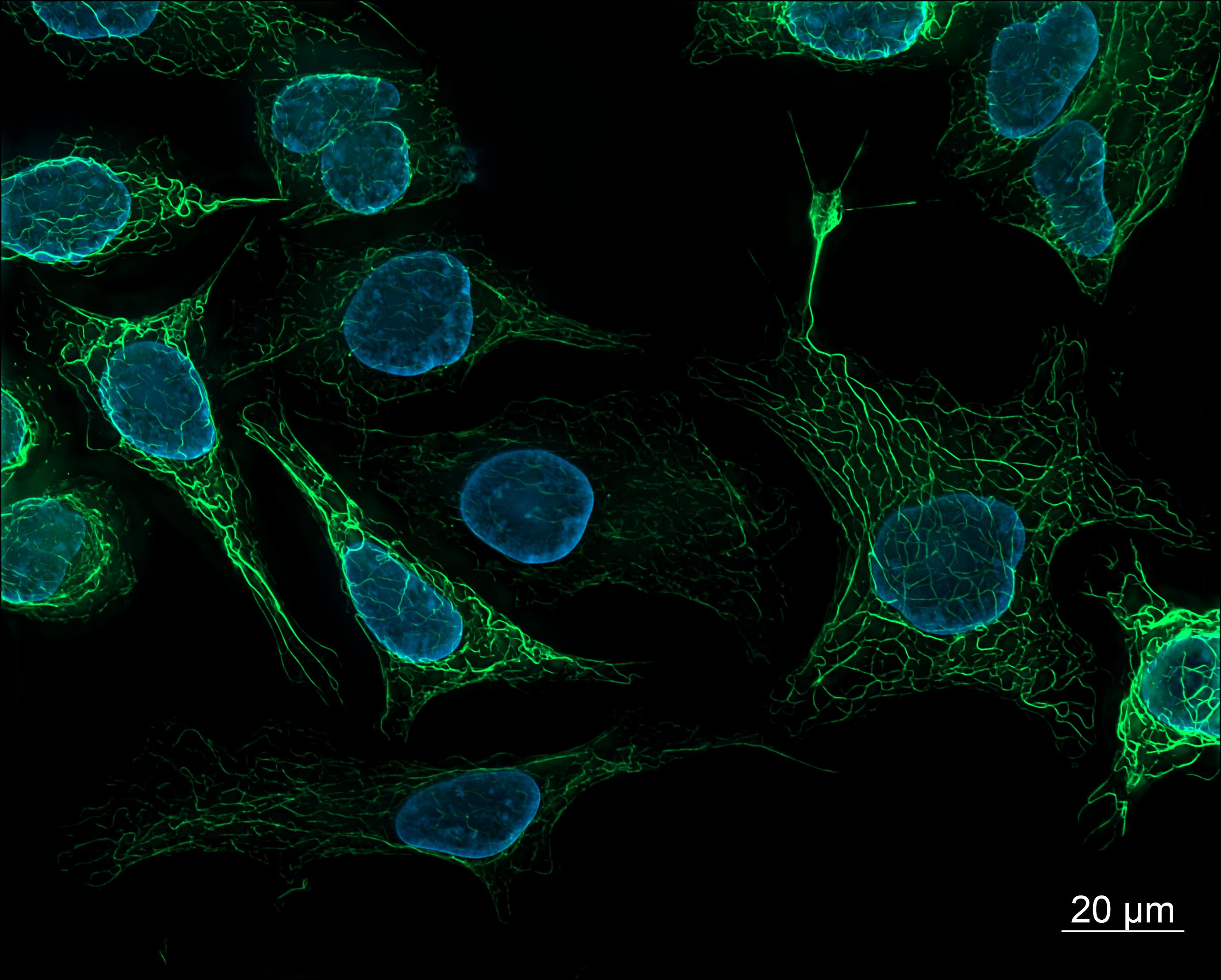
File:SK8-18-2 human derived cells, fluorescence microscopy  (29942101073).jpg - Wikimedia Commons
File:SK8-18-2 human derived cells, fluorescence microscopy (29942101073).jpg - Wikimedia Commons

Flexible warped nano-graphene shows promise for fluorescent cell imaging

Cell Explorer™ Live Cell Labeling Kit *Green Fluorescence* | AAT Bioquest

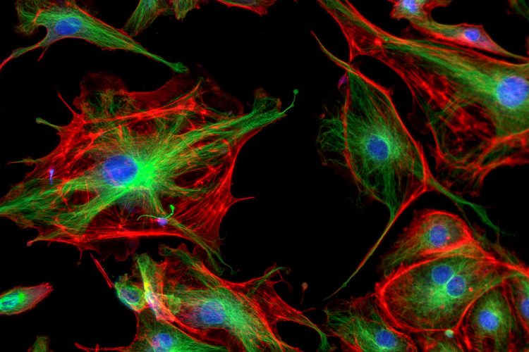
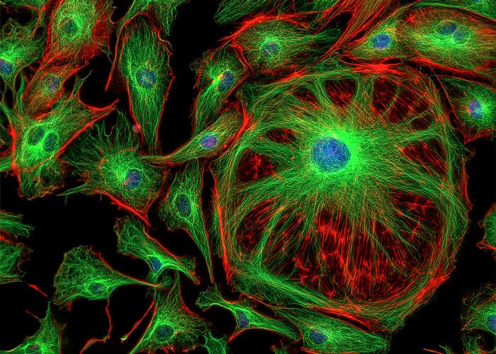
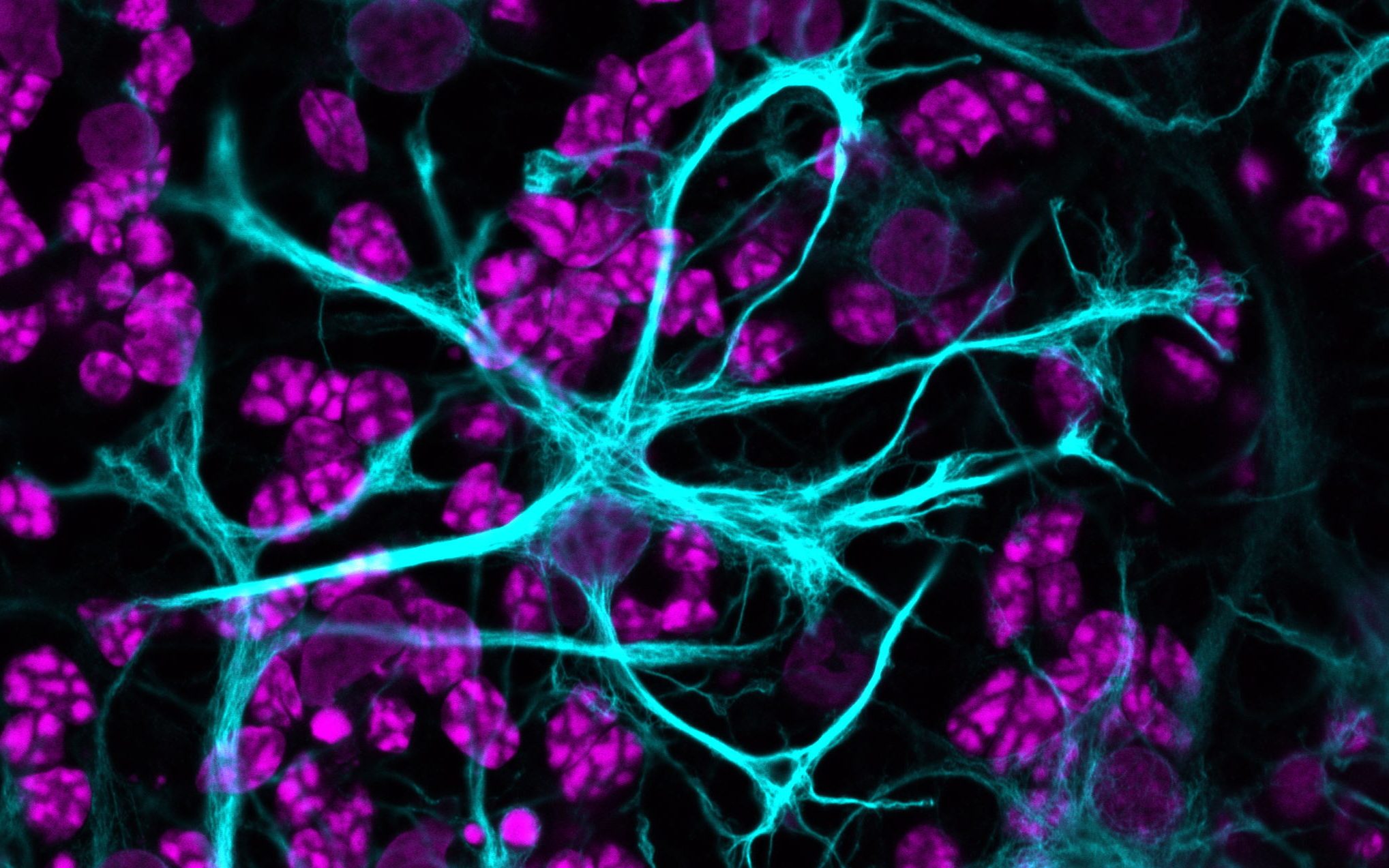
Fluorescence Image of Human Cells with Cytoskeletal Microtubules and DNA  Stock Image - Image of magenta, fluorescence: 106699101
Fluorescence Image of Human Cells with Cytoskeletal Microtubules and DNA Stock Image - Image of magenta, fluorescence: 106699101

Fluorescent microscopy - LNF Wiki

Improving how we image cells using carbon dots - Advanced Science News

Using QuPath to calculate cells with different stain in fluorescence -  Image Analysis - Image.sc Forum
Using QuPath to calculate cells with different stain in fluorescence - Image Analysis - Image.sc Forum
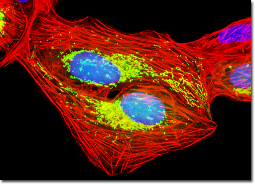
Lower background fluorescence in live cell imaging - Tebubio's blog

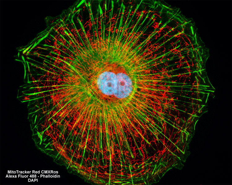
Molecular Expressions Microscopy Primer: Specialized Microscopy Techniques  - Fluorescence Digital Image Gallery - Human Bone Osteosarcoma Cells (U-2  OS)
Molecular Expressions Microscopy Primer: Specialized Microscopy Techniques - Fluorescence Digital Image Gallery - Human Bone Osteosarcoma Cells (U-2 OS)
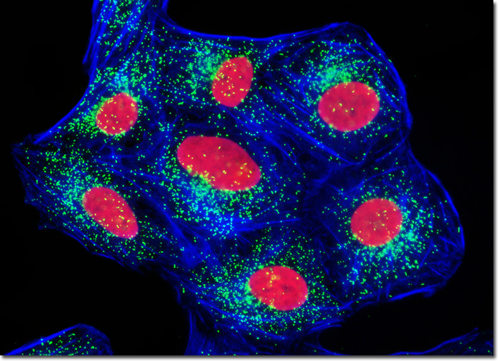
6 Tips for Fluorescence Live Cell Imaging | Olympus LS

ZEISS Microscopy Online Campus | Fluorescence Image Gallery | Adherent Cells

Image of the Day | The Scientist Magazine® | Microscopic photography, Stem  cells, Image of the day
Image of the Day | The Scientist Magazine® | Microscopic photography, Stem cells, Image of the day

Choosing Fluorescent Reagents for Every Live Cell Application | The  Scientist Magazine®
Choosing Fluorescent Reagents for Every Live Cell Application | The Scientist Magazine®

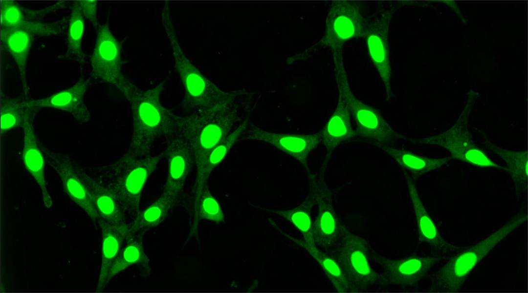
Fluorescent cells grown using CMOS image sensors

Fluorescent Cell Lines – Cells Online

Molecular Expressions Microscopy Primer: Specialized Microscopy Techniques  - Fluorescence Digital Image Gallery - Human Bone Osteosarcoma Cells (U-2  OS)
Molecular Expressions Microscopy Primer: Specialized Microscopy Techniques - Fluorescence Digital Image Gallery - Human Bone Osteosarcoma Cells (U-2 OS)
How to detect Singlet Oxygen by live cell imaging? - Tebubio's blog

3.3D: Fluorescence Microscopy - Biology LibreTexts

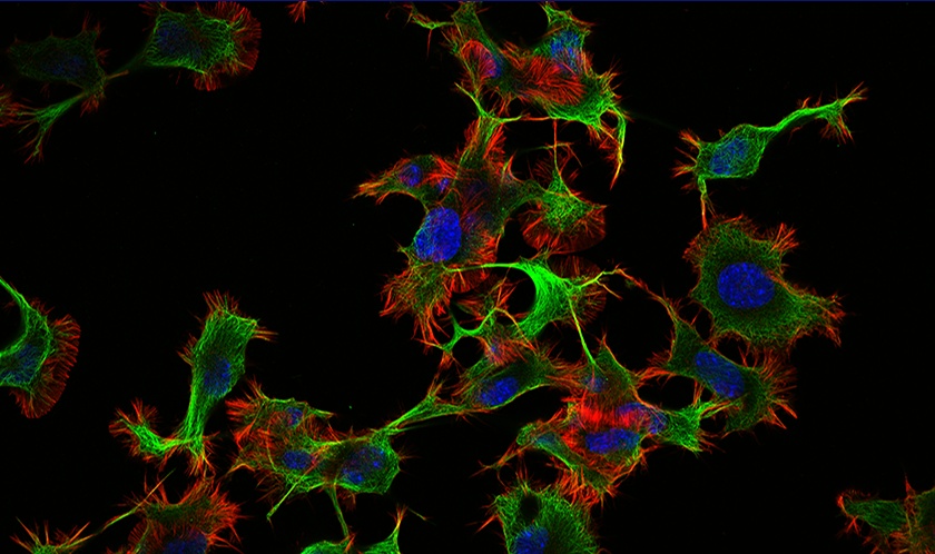
Image of the Day: Fluorescent Cells | The Scientist Magazine®

Cell Navigator® Mitochondrion Staining Kit *NIR Fluorescence* - Stratech

Cell painting, Phenotypic and Morphological Profiling | Molecular Devices

Fluorescent labeling of proteins inside the living cell with high... |  Download Scientific Diagram
Fluorescent labeling of proteins inside the living cell with high... | Download Scientific Diagram

Colours in the dark: fluorescence microscopy for the classroom – Science in  School
Colours in the dark: fluorescence microscopy for the classroom – Science in School

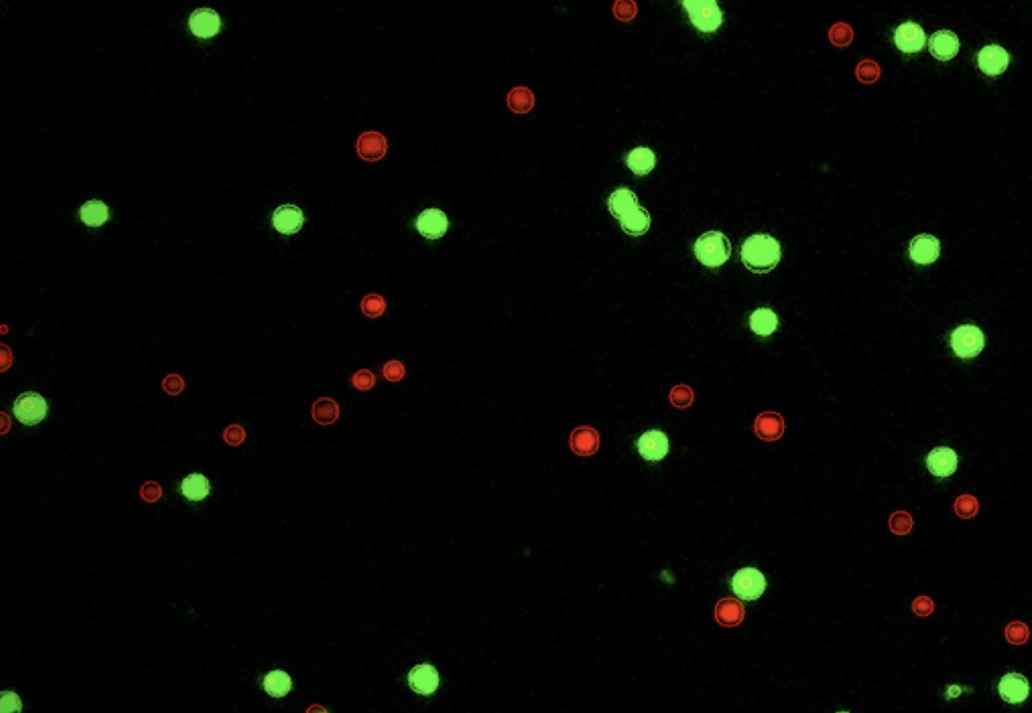
Quantifying Fluorescent Cells in Mammalian Cell Tissue Culture

Automated Fluorescence Cell Counting – Logos Biosystems | Advanced Imaging  Solutions for Research Excellence
Automated Fluorescence Cell Counting – Logos Biosystems | Advanced Imaging Solutions for Research Excellence